
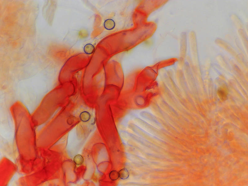

Red-tinted tissues of Ascomycete fungus Hyaloscypha britannica under a microscope
💾 Download Full Size
Highest-resolution version of the photo
|
📷 Shooting data
Sat 7 May 2022 14:30:14 - USB Camera
📊 Histogram
![Histogram Preview]()

▦ Current Album

|
Album 3148: Sam Houston Forest, Texas, May 7, 2022
The image shows three red-tinted fungal hyphae with a few yellowish spores attached to them. The red tint is due to the presence of a substance like iodine or potassium hydroxide, which stains the tissues of certain fungi.
In the background, there are some light-colored structures that resemble other fungal hyphae or possibly plant cells.
The image was taken under a microscope on May 7, 2022. The specimen is likely from an Ascomycete fungus called Hyaloscypha britannica. – AI vision


