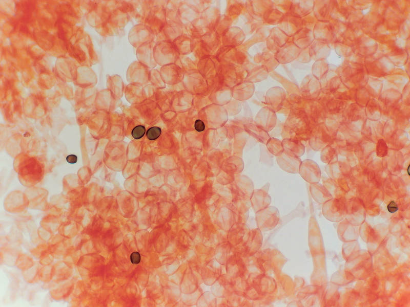

Gill tissue and spores of a white brittle gilled Hemimycena(?) mushroom under low magnification, collected on Stubblefield section of Lone Star hiking trail in Sam Houston National Forest. Texas


Album 3192: Sam Houston Forest, Texas, July 16 - 17, 2022
The image presents a microscopic view of Gill tissue and spores from an unidentified Hemimycena(?) mushroom, collected on Stubblefield section of Lone Star hiking trail in Sam Houston National Forest, Texas, July 16, 2022.
* **Microscopic View:**
* The image displays a low-magnification microscopic view of the gill tissue and spores.
* The background is white, with numerous small, circular structures visible throughout.
* The circular structures are likely individual cells or cell clusters within the gill tissue.
* **Spore Characteristics:**
* Several dark brown or black dots are scattered throughout the image.
* These dots represent spores, which are reproductive structures produced by fungi for dispersal and propagation.
* Spores can be spherical, oval, cylindrical, or other shapes depending on the fungal species.
* **Gill Tissue Appearance:**
* The gill tissue appears as a dense network of fine, branching filaments.
* These filaments are composed of mycelium, which is the vegetative part of a fungus's body.
* Mycelium grows by extending its hyphae, tiny branching fibers that make up the fungal structure.
* **Coloration and Staining:**
* The gill tissue and spores are stained with a reddish-orange dye, which highlights their morphology.
* The staining technique used is likely lacto-phenol cotton blue or similar, commonly employed for examining fungi at low magnification.
In summary, the image shows a microscopic view of gill tissue and spores from an unidentified Hemimycena(?) mushroom. The gill tissue appears as a dense network of fine filaments, while the spores are visible as dark brown or black dots scattered throughout the image. The staining technique used highlights the morphology of these structures, providing valuable information for further identification and study of this fungal species. – AI vision
🇪🇸 Descripción en español / Spanish Description
Tejido de las branquias y esporas de un hongo Hemimycena (?) blando con garras blancas, capturado a baja magnificación, recogido en la sección Stubblefield del sendero Lone Star en el bosque nacional Sam Houston. Texas, 16 de julio de 2022. La imagen presenta una vista microscópica del tejido de las branquias y esporas de un hongo Hemimycena (?) no identificado, recogido en la sección Stubblefield del sendero Lone Star en el bosque nacional Sam Houston, Texas, el 16 de julio de 2022. * **Vista microscópica:** * La imagen muestra una vista microscópica a baja magnificación del tejido de las branquias y esporas. * El fondo es blanco, con numerosas estructuras circulares pequeñas visibles en toda su extensión. * Las estructuras circulares son probablemente células individuales o agrupaciones celulares dentro del tejido de las branquias. * **Características de las esporas:** * Varios puntos marrones oscuros o negros se dispersan por toda la imagen. * Estos puntos representan esporas, que son estructuras reproductivas producidas por hongos para su dispersión y propagación. * Las esporas pueden ser esféricas, ovaladas, cilíndricas u otras formas dependiendo de la especie fúngica. * **Apariencia del tejido de las branquias:** * El tejido de las branquias aparece como una densa red de filamentos finos y ramificados. * Estos filamentos están compuestos por micelio, que es la parte vegetativa del cuerpo de los hongos. * El micelio crece extendiendo sus hifas, pequeñas fibras ramificadas que constituyen la estructura fúngica. * **Coloración y tinción:** * Las esporas y el tejido de las branquias se tiñeron con un colorante rojo anaranjado, lo que resalta su morfología. * La técnica de tinción utilizada es probablemente la lacto-fenol azul de algodón o similar, empleada comúnmente para examinar hongos a baja magnificación. En resumen, la imagen muestra una vista microscópica del tejido de las branquias y esporas de un hongo Hemimycena (?) blando con garras blancas. El tejido de las branquias aparece como una densa red de filamentos finos, mientras que las esporas son visibles como puntos oscuros o marrones dispersados por toda la imagen. La técnica de tinción utilizada resalta la morfología de estas estructuras, proporcionando información valiosa para la identificación y el estudio adicionales de esta especie fúngica. – Spanish translation