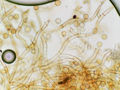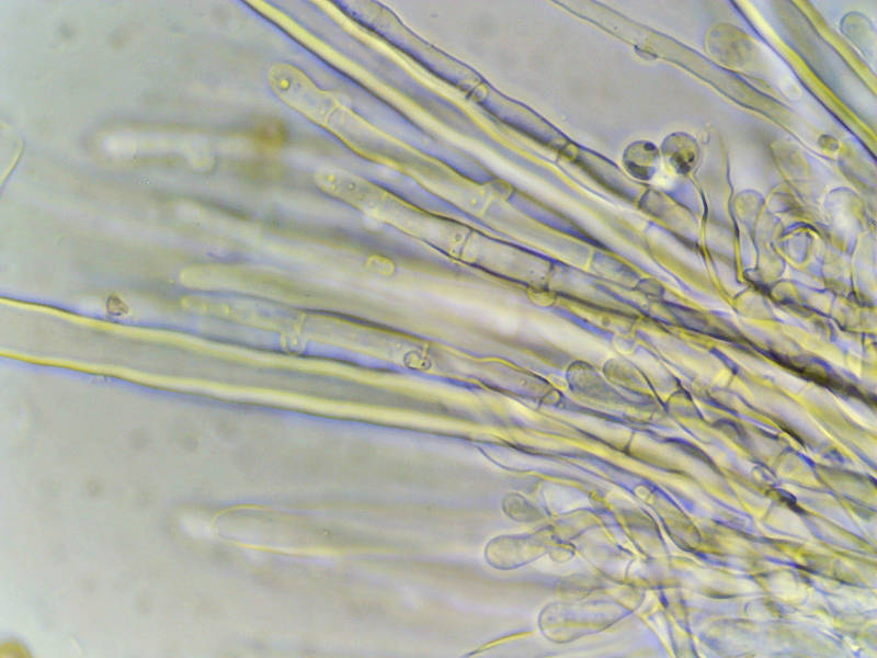

Tissues and conidia(?) of bluish Tomentella(?) fungus, from the same oak log as brown Botryobasidium and common poria (in KOH), collected on Kiwanis Nature Trail. College Station, Texas


Album 3401: College Station, Texas, May 1, 2023
The image shows a microscopic view of fungal hyphae in a preparation stained with a solution called potassium hydroxide (KOH). The background is mostly transparent and appears greyish-blue due to the staining effect of the KOH on the hyphae.
**Key Features:**
* **Hyphae:** These are the long, thin threads that make up the fungal mycelium. They have a rounded tip at one end and taper towards the other.
* **Conidia (spores):** Several small, spherical structures can be seen attached to the hyphae. These are likely conidial spores, which are produced by some fungi as a means of reproduction.
**Significance:**
The presence of these features suggests that this sample is from a fungus in the order Polyporales. The KOH staining has highlighted the fungal structures, allowing for easy observation under a microscope.
**Conclusion:**
Based on the microscopic image and the context provided, it appears that the image shows a Tomentella fungus with conidial spores attached to its hyphae. – AI vision
🇪🇸 Descripción en español / Spanish Description
Los tejidos y los conidios (?) del hongo Tomentella(?) bluish, del mismo tronco de roble que el Botryobasidium marrón y la poria común (en KOH), recolectados en el sendero Kiwanis Nature Trail. College Station, Texas, 1 de mayo de 2023. La imagen muestra una vista microscópica de las hifas fúngicas en una preparación teñida con una solución llamada hidróxido de potasio (KOH). El fondo es en su mayoría transparente y aparece grisáceo debido al efecto de tinción del KOH sobre las hifas. **Características clave:** * **Hifas:** Son los hilos largos y delgados que forman el micelio fúngico. Tienen una punta redondeada en un extremo y se estrechan hacia el otro. * **Conidios (esporas):** Se pueden ver varias estructuras esféricas pequeñas unidas a las hifas. Estos son probablemente conidiósporos, que producen algunos hongos como medio de reproducción. **Importancia:** La presencia de estas características sugiere que esta muestra pertenece a un hongo del orden Polyporales. El teñido KOH ha resaltado las estructuras fúngicas, lo que permite una fácil observación bajo el microscopio. **Conclusión:** Basándose en la imagen microscópica y el contexto proporcionado, parece que la imagen muestra un hongo Tomentella con esporas conidiales unidas a sus hifas. – Spanish translation