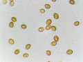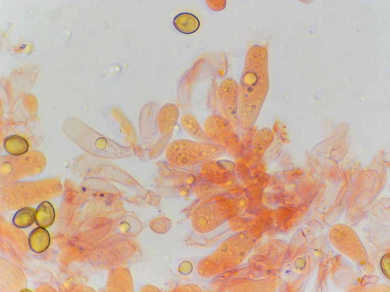

Basidia and spores (in Congo Red) of tiny pleurotoid mushrooms Simocybe amara(?) from a fallen trunk of Japanese privet, collected in Hensel Park. College Station, Texas


Album 3408: College Station, Texas, May 8, 2023
The image depicts a microscopic view of fungal spores and basidia, likely from the species Simocybe amara. The spores are small, oval-shaped structures that range in color from light brown to dark yellowish-brown. They have smooth surfaces with no visible features.
**Basidium**
In the center of the image is a cluster of elongated cells called basidia. Basidia are responsible for producing spores in fungi and are typically found at the base of gills or pores on the underside of a mushroom cap. The basidia appear to be slightly curved, with some having small droplets or bubbles attached to their surfaces.
**Spore Formation**
The image suggests that the spores are being produced by the basidia, which is consistent with the reproductive cycle of fungi. When conditions are favorable, such as high humidity and warmth, the basidia will release their spores into the air, allowing them to disperse and potentially colonize new areas.
**Microscopic Details**
The image provides a detailed view of the microscopic features of the fungal cells, including the shape, size, and coloration of the spores. The basidia are also clearly visible, with their characteristic elongated shape and curved appearance. Overall, the image offers a fascinating glimpse into the microscopic world of fungi and highlights the intricate details that underlie their reproductive processes.
*Answer*: Microscopic view of Simocybe amara spores and basidia. – AI vision
🇪🇸 Descripción en español / Spanish Description
Vista microscópica de esporas y basidios del minúsculo hongo pleurotoide Simocybe amara (?) procedente del tronco caído de un laurel japonés, recogido en Hensel Park. College Station, Texas, 8 de mayo de 2023. La imagen representa una vista microscópica de esporas fúngicas y basidios, probablemente de la especie Simocybe amara. Las esporas son estructuras ovaladas pequeñas cuyo color varía entre marrón claro y marrón amarillento oscuro. Tienen superficies lisas sin características visibles. **Basidios** En el centro de la imagen hay un grupo de células alargadas llamadas basidios. Los basidios son responsables de producir esporas en los hongos y se encuentran normalmente en la base de las láminas o poros en la cara inferior del sombrero del hongo. Los basidios parecen estar ligeramente curvados, algunos con pequeñas gotas o burbujas unidas a sus superficies. **Formación de esporas** La imagen sugiere que las esporas están siendo producidas por los basidios, lo que es coherente con el ciclo reproductivo de los hongos. Cuando las condiciones son favorables, como humedad y calor elevados, los basidios liberan sus esporas al aire, permitiendo que éstas se dispersen y potencialmente colonicen nuevas áreas. **Detalles microscópicos** La imagen ofrece una vista detallada de las características microscópicas de las células fúngicas, incluyendo la forma, tamaño y coloración de las esporas. Los basidios también son claramente visibles con su característica forma alargada y apariencia curvada. En general, la imagen ofrece una fascinante visión del mundo microscópico de los hongos y destaca los intrincados detalles que subyacen a sus procesos reproductivos. *Respuesta*: Vista microscópica de esporas y basidios de Simocybe amara. – Spanish translation