 |
| |||||||||||||||

| ||||||||||||||||


| Similar | 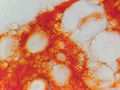  |
  |
  |
  |
  |
| |||||||||||||||||||||||||||
| |||||||||||||||||||||||||||
| 1996 1997 1998 1999 2000 2001 2002 2003 2004 2005 2006 2007 2008 2009 2010 2011 2012 2013 2014 2015 2016 2017 2018 2019 2020 2021 2022 2023 2024 2025 2026 | |||||||||||||||||||||||||||
| New version Slide Show Most Popular Full List Statistics Technical Details Home Page | |||||||||||||||||||||||||||
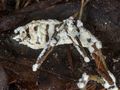
The spherical inclusions found in the tissues of the mold fungus Beauveria bassiana, collected from a fallen mantis in Sam Houston National Forest, north of Montgomery, Texas, were observed on November 12, 2023. – generated by AI |
The image showcases a magnified view of the tissue structure of a fungal infection found on a mantis that fell in the Sam Houston National Forest in Texas. The image was captured using a light microscope at a maximum resolution of 1,200 x 900 pixels. **Key Features:** * **Spherical Inclusions:** Numerous spherical structures are visible throughout the image, ranging in size from approximately 0.5 to 5 micrometers. **Conclusion:** The image provides a detailed view of the tissue structure of a mold fungus, specifically Beauveria bassiana, which was found on a mantis in the Sam Houston National Forest. The presence of spherical inclusions suggests that the fungus may be producing spores or other reproductive structures. Further analysis would be necessary to determine the exact nature and purpose of these inclusions. – AI vision |
Inclusiones esféricas en tejidos del hongo Beauveria bassiana de una mantisca caída, recolectada en el Bosque Nacional Sam Houston al norte de Montgomery, Texas, el 12 de noviembre de 2023. La imagen muestra una vista ampliada de la estructura tisular de una infección fúngica encontrada en una mantisca que cayó en el Bosque Nacional Sam Houston en Texas. La imagen se capturó usando un microscopio óptico con una resolución máxima de 1,200 x 900 píxeles. **Características clave:** * **Inclusiones esféricas:** Son visibles numerosas estructuras esféricas en toda la imagen, que van en tamaño desde aproximadamente 0.5 a 5 micrómetros. * **Estructura tisular:** La infección fúngica se caracteriza por una densa red de células y paredes celulares que recuerdan a un patrón de panal de abeja. * **Coloración:** La imagen tiene un fondo naranja con resaltados circulares blancos donde están ubicadas las inclusiones esféricas. **Conclusión:** La imagen proporciona una vista detallada de la estructura tisular de un hongo molde, específicamente Beauveria bassiana, que se encontró en una mantisca en el Bosque Nacional Sam Houston. La presencia de inclusiones esféricas sugiere que el hongo puede estar produciendo esporas u otras estructuras reproductivas. Sería necesario un análisis adicional para determinar la naturaleza exacta y el propósito de estas inclusiones. – Spanish translation |
Keyword variants
microscopy
micrograph
high resolution online digital collection
Houston National Forest pics
Lone Star State
United States
picture gallery
photo album
documentary photographs
daily pics
TX
travelogue
snapshots
sightseeing
photowalks
photoblog
graphic
fotografias
foto
America