 |
| |||||||||||||||

| ||||||||||||||||


| Similar |   |
  |
  |
  |
  |
| |||||||||||||||||||||||||||
| |||||||||||||||||||||||||||
| 1996 1997 1998 1999 2000 2001 2002 2003 2004 2005 2006 2007 2008 2009 2010 2011 2012 2013 2014 2015 2016 2017 2018 2019 2020 2021 2022 2023 2024 2025 2026 | |||||||||||||||||||||||||||
| New version Slide Show Most Popular Full List Statistics Technical Details Home Page | |||||||||||||||||||||||||||
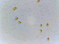
Thin white corticioid fungi spores were collected from hardwood trees lacking bark in the Sam Houston National Forest of Texas on November 20, 2023. The collection was made a day prior. – generated by AI |
The image shows a microscopic view of spores of a thin white corticioid fungus on a slide. The spores are numerous and randomly distributed within the field of view, appearing as small, roundish or oval-shaped structures with smooth edges. **Key Features:** * **Spore Distribution:** The spores are densely packed but not overlapping, giving an impression that they could be counted. **Conclusion:** The image likely represents a species of corticioid fungus, which belongs to a larger group of fungi called Corticiales. These fungi are characterized by their thin, crust-like growths on dead wood and have been traditionally classified based on morphological characteristics such as spore size and shape. However, without further information or context about the specimen's collection site, time of year, and any additional microscopic features like hyphae structures, it is difficult to provide a more specific identification beyond noting that the sample appears to be a corticioid fungus. **Answer:** Corticioid Fungus. – AI vision |
Translate the following text from English to Spanish: Las esporas de un hongo corticío blanco delgado de madera dura sin corteza, recogido un día antes en el Bosque Nacional Sam Houston. Texas, 20 de noviembre de 2023. La imagen muestra una vista microscópica de esporas de un hongo corticío blanco delgado en un portaobjetos. Las esporas son numerosas y se distribuyen aleatoriamente dentro del campo de visión, apareciendo como estructuras redondeadas ovaladas pequeñas con bordes lisos. **Características clave:** * **Distribución de esporas:** Las esporas están densamente empaquetadas pero no se solapan, dando la impresión de que podrían contarse. * **Tamaño y forma:** Las esporas varían ligeramente en tamaño pero generalmente parecen ser de unos 10-15 micrómetros de diámetro. Sus formas varían desde redondeadas a ovaladas, apareciendo algunas más alargadas que otras. * **Coloración:** Las esporas son amarillo-parduzcas uniformes, lo que indica que no han sido teñidas ni coloreadas para el examen microscópico. * **Fondo:** El fondo de la imagen tiene un color blanco grisáceo claro, típico de los portaobjetos de microscopio. Proporciona un contraste neutro con las esporas amarillo-parduzcas, lo que hace que destaquen claramente. **Conclusión:** La imagen representa probablemente una especie de hongo corticío, que pertenece a un grupo más grande de hongos llamado Corticiales. Estos hongos se caracterizan por sus crecimientos similares a costra sobre madera muerta y se han clasificado tradicionalmente en función de características morfológicas como el tamaño y la forma de las esporas. Sin embargo, sin más información o contexto sobre el sitio de recolección del espécimen, la época del año y cualquier otra característica microscópica adicional como estructuras de hifas, es difícil proporcionar una identificación más específica. – Spanish translation |
Keyword variants
microscopy
micrograph
high resolution online digital collection
Houston National Forest photo
Lone Star State
United States
picture gallery
photo album
documentary photographs
daily pics
TX
travelogue
snapshots
sightseeing
photowalks
photoblog
graphic
fotografias
foto
America