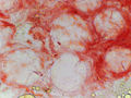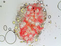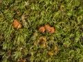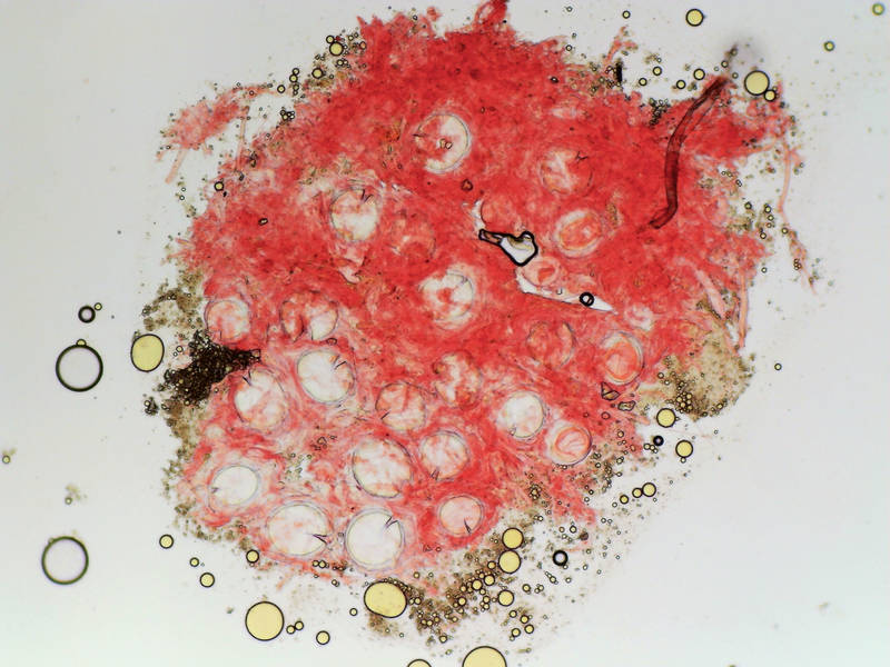

Tissues (in Congo Red) of orange sclerotia of some fungus under a microscope at low magnification, collected north from Trailhead No. 6 in Sam Houston National Forest. Texas


Album 3452: College Station - Sam Houston Forest, Texas, December 30 - 31, 2023
The image shows a fungal growth, likely an orange sclerotia, characterized by its vibrant red color and spherical shape. The sclerotia appears to be composed of numerous small, round cells that are densely packed together. These cells are stained with Congo Red, which is commonly used to visualize the presence of certain types of fungi.
**Key Observations:**
* **Color:** The fungal growth exhibits a distinctive orange-red color due to the staining with Congo Red.
* **Shape:** The sclerotia has a spherical shape, indicating that it is likely a type of fungus that grows in compact, rounded masses.
* **Cell Structure:** The cells within the sclerotia are small and round, suggesting that they may be spores or other reproductive structures.
**Conclusion:**
The image suggests that the fungal growth is an orange sclerotia, possibly belonging to a species of Armillaria mellea. This conclusion is based on the characteristic coloration and cell structure observed in the image. However, further examination and analysis would be necessary to confirm the identity of the fungus with certainty.
*Answer*: The image shows a fungal growth, likely an orange sclerotia, characterized by its vibrant red color and spherical shape, stained with Congo Red to visualize the presence of certain types of fungi. – AI vision
🇪🇸 Descripción en español / Spanish Description
Muestras (en rojo Congo) de esclerocios de color naranja de cierto hongo bajo un microscopio con baja ampliación, recogidas al norte del punto de partida número 6 en el bosque nacional Sam Houston, Texas. 31 de diciembre de 2023. La imagen muestra un crecimiento fúngico, probablemente un esclerocio naranja, caracterizado por su color rojo vibrante y su forma esférica. El esclerocio parece estar compuesto por numerosas células pequeñas y redondas que están densamente empaquetadas juntas. Estas células se tiñen con Rojo Congo, lo cual se usa comúnmente para visualizar la presencia de ciertos tipos de hongos. **Observaciones clave:** * **Color:** El crecimiento fúngico exhibe un distintivo color naranja-rojo debido a la tinción con Rojo Congo. * **Forma:** El esclerocio tiene una forma esférica, lo que indica que probablemente sea un tipo de hongo que crece en masas compactas y redondeadas. * **Estructura celular:** Las células dentro del esclerocio son pequeñas y redondas, sugiriendo que podrían ser esporas u otras estructuras reproductivas. **Conclusión:** La imagen sugiere que el crecimiento fúngico es un esclerocio naranja, posiblemente perteneciente a una especie de Armillaria mellea. Esta conclusión se basa en la característica coloración y estructura celular observadas en la imagen. Sin embargo, sería necesario un examen y análisis adicionales para confirmar la identidad del hongo con certeza. *Respuesta*: La imagen muestra un crecimiento fúngico, probablemente un esclerocio naranja, caracterizado por su color rojo vibrante y su forma esférica, teñido con Rojo Congo para visualizar la presencia de ciertos tipos de hongos. – Spanish translation