 |
| |||||||||||||||

| ||||||||||||||||


| Similar |   |
  |
  |
  |
  |
| |||||||||||||||||||||||||||
| |||||||||||||||||||||||||||
| 1996 1997 1998 1999 2000 2001 2002 2003 2004 2005 2006 2007 2008 2009 2010 2011 2012 2013 2014 2015 2016 2017 2018 2019 2020 2021 2022 2023 2024 2025 2026 | |||||||||||||||||||||||||||
| New version Slide Show Most Popular Full List Statistics Technical Details Home Page | |||||||||||||||||||||||||||
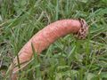
On January 3, 2024, I collected stalked lattice stinkhorn mushroom (Lysurus periphragmoides) spores and tissues from a lawn in Hensel Park, College Station, Texas. The specimens were preserved in Congo Red and feature intricate lattice-like structures on their surfaces. These unique features make them an interesting subject for study and observation. – generated by AI |
The image shows microscopic slides of stalked Lattice Stinkhorn mushroom (Lysurus periphragmoides) spores and tissues that were collected from a lawn in Hensel Park on January 3, 2024. **Spore Characteristics:** * The spores are oval to elliptical in shape. **Tissue Characteristics:** * The tissues appear as a network of fibers with some granular material. **Overall Appearance:** * The microscopic image displays two distinct types of structures: spores and tissues. |
Esperas y tejidos (en rojo Congo) de la seta huela a rejilla con pedúnculo (Lysurus periphragmoides) de un césped, recogida en un prado de Hensel Park. College Station, Texas, 3 de enero de 2024. La imagen muestra portaobjetos microscópicos de esporas y tejidos de seta huela a rejilla con pedúnculo (Lysurus periphragmoides) que se recogieron en un prado de Hensel Park el 3 de enero de 2024. **Características de las esporas:** * Las esporas tienen una forma de óvalo a elipse. * Tienen una superficie lisa con algunas irregularidades cerca de los extremos. * El color de las esporas varía desde amarillo pálido hasta marrón claro, y su tamaño también varía, pero la mayoría miden entre 2,5-3,5 micrómetros por 1,2-1,8 micrómetros. **Características de los tejidos:** * Los tejidos aparecen como una red de fibras con algo de material granulado. * Se tiñen en rojo con el colorante Congo Red. * Las fibras parecen estar un poco retorcidas y curvas, lo que les da un aspecto ondulado. * Algunas áreas muestran un teñido más oscuro debido a la presencia de pigmentación más intensa. **Apariencia general:** * La imagen microscópica muestra dos tipos distintos de estructuras: esporas y tejidos. * Las esporas están dispersas por todo el campo de visión, mientras que los tejidos aparecen como una red de fibras en algunas áreas y como aglomeraciones aisladas de material granulado en otras. – Spanish translation |
Keyword variants
microscopy
micrograph
high resolution online digital collection
Berlin on the Brazos
Lone Star State
Hensel Park image
United States
picture gallery
photo album
documentary photographs
daily pics
conservative town
Brazos County
TX
travelogue
snapshots
sightseeing
photowalks
photoblog
graphic
fungus
fungi
fotografias
foto
Bryan
America
Aggieland