 |
| |||||||||||||||||

| ||||||||||||||||||


| Similar | 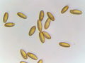  |
  |
  |
  |
  |
| |||||||||||||||||||||||||||||
| |||||||||||||||||||||||||||||
| 1996 1997 1998 1999 2000 2001 2002 2003 2004 2005 2006 2007 2008 2009 2010 2011 2012 2013 2014 2015 2016 2017 2018 2019 2020 2021 2022 2023 2024 2025 2026 | |||||||||||||||||||||||||||||
| New version Slide Show Most Popular Full List Statistics Technical Details Home Page | |||||||||||||||||||||||||||||
The image presents a microscopic view of spores from the red cracking bolete mushroom, collected on July 23rd and observed on August 22nd at the Polytechnic Institute in St. Petersburg, Russia. The photograph was taken using potassium hydroxide (KOH) as a mounting medium. **Image Description:** * **Background:** The background is white. **Key Observations:** * The image provides a detailed view of the spore morphology, including their shape, size, and coloration. **Conclusion:** The image offers a detailed look at the spores of the red cracking bolete mushroom, providing valuable information for those interested in mycology and fungal identification. – AI vision |
Споры (в КОН) красного трещинового подберезовика (Xerocomellus chrysenteron), собранные ранее 23 июля 2024 г. (фото 3575-22) в Садах Политехнического института, Санкт-Петербург, Россия, 22 августа 2024 года. На изображении представлен микроскопический вид спор красного трещинового подберезовика, собранных 23 июля и наблюдаемых 22 августа в Политехническом институте Санкт-Петербурга. Фотография сделана с использованием поташа (КОН) в качестве среды для монтирования. **Описание изображения:** * **Фон:** Фон белый. * **Споры:** Овальные желтовато-коричневые споры с гладкой поверхностью. Имеют неправильную форму и различаются по размеру, достигая длины приблизительно от 15 до 40 микрометров. * **Среда для монтирования:** Споры были монтированы с использованием КОН, что является распространенной техникой подготовки грибных образцов для микроскопии. * **Ключевые наблюдения:** * На изображении представлен детальный вид морфологии спор, включая их форму, размер и окраску. * Использование КОН в качестве среды для монтирования позволяет четко визуализировать особенности поверхности спор. * Фотография сделана в микроскопическом масштабе, что говорит о ее предполагаемом использовании в научных или образовательных целях. **Заключение:** Изображение дает подробное представление о спорах красного трещинового подберезовика, предоставляя ценную информацию для людей, интересующихся микологией и идентификацией грибов. – Russian translation |
Keyword variants
microscopy
micrograph
high resolution online digital collection
St. Pete's
St. Petersburg pix
Soviet Union
Sankt Peterburg
Saint Petersburg
Saint-Petersbourg
Russian Federation
Rossiya foto
picture gallery
photo album
Leningrad fotosnimki
former USSR
documentary photographs
daily pics
Venemaa
Venaja
travelogue
SPb
snimki
snapshots
sightseeing
Ryssland
Rwsia
Ruthenia
Rusya
Rusujo
Russland
Russja
Russie
Russi
Ruslando
Rusland
Rusko
Rusio
Rusija
Rusii
Rusia
Roushie
Rosya
Rosland
Rosia
Roshia
Raseja
Piter
Pietari
photowalks
photoblog
Petrograd
Orussiya
Oroszorszag
Lenjingrad
institue
graphic
fungus
fungi
fotografias
fotki
Errusia