 |
| |||||||||||||||

| ||||||||||||||||


| Similar |   |
  |
  |
  |
  |
| |||||||||||||||||||||||||||
| |||||||||||||||||||||||||||
| 1996 1997 1998 1999 2000 2001 2002 2003 2004 2005 2006 2007 2008 2009 2010 2011 2012 2013 2014 2015 2016 2017 2018 2019 2020 2021 2022 2023 2024 2025 2026 | |||||||||||||||||||||||||||
| New version Slide Show Most Popular Full List Statistics Technical Details Home Page | |||||||||||||||||||||||||||
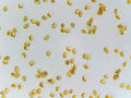
The image shows a microscopic view of spores, specifically those of the Tyromyces mushroom. The spores are yellowish-brown and ovoid in shape, with a smooth surface. They vary in size but most appear to be about 5-7 micrometers long. The background of the image is light gray, which suggests that it was taken using a microscope with a phase contrast or differential interference contrast (DIC) objective. This technique enhances the visibility of small details such as cell membranes and organelles by highlighting differences in refractive index between different parts of the sample. In this case, the yellowish-brown coloration of the spores indicates that they are likely to be basidiospores, which are produced by the basidia (spore-bearing cells) of fungi. The presence of these spores suggests that the mushroom is a member of the genus Tyromyces, which includes several species that grow on trees and other woody plants. Overall, the image provides valuable information about the morphology and structure of Tyromyces spores, which can be useful for identifying and characterizing this fungus. It also highlights the importance of microscopy in understanding the biology and ecology of fungi, particularly those that play a crucial role in decomposing organic matter and recycling nutrients in ecosystems. – AI vision |
Las esporas (en lugol) del hongo Tyromyces de la base del olmo en una zona húmeda y barrosa, recogidas en la sección de Caney Creek del Lone Star Hiking Trail en el Bosque Nacional Sam Houston al norte de Montgomery. Texas, 28 de septiembre de 2024. La imagen muestra una vista microscópica de esporas, específicamente las del hongo Tyromyces. Las esporas son amarillentas-marrón y de forma ovoide, con una superficie lisa. Varios en tamaño pero la mayoría parecen ser de aproximadamente 5-7 micrómetros de largo. El fondo de la imagen es gris claro, lo que sugiere que se tomó usando un microscopio con un objetivo de contraste de fase o diferencial de interferencia (DIC). Esta técnica mejora la visibilidad de pequeños detalles como membranas celulares y orgánulos resaltando las diferencias en el índice de refracción entre diferentes partes de la muestra. En este caso, la coloración amarillenta-marrón de las esporas indica que probablemente sean basidiosporas, que son producidas por los basidios (células portadoras de esporas) de hongos. La presencia de estas esporas sugiere que el hongo es un miembro del género Tyromyces, que incluye varias especies que crecen en árboles y otras plantas leñosas. En general, la imagen proporciona información valiosa sobre la morfología y estructura de las esporas de Tyromyces, lo que puede ser útil para identificar y caracterizar este hongo. También destaca la importancia de la microscopía en comprender la biología y ecología de los hongos, particularmente aquellos que desempeñan un papel crucial en la descomposición de la materia orgánica y el reciclaje de nutrientes en los ecosistemas. – Spanish translation |
Keyword variants
microscopy
micrograph
high resolution online digital collection
Star Hiking Trail pictures
Houston National Forest images
Lone Star State
United States
picture gallery
photo album
documentary photographs
daily pics
TX
travelogue
snapshots
sightseeing
photowalks
photoblog
graphic
fungus
fungi
fotografias
foto
America