 |
| |||||||||||||||

| ||||||||||||||||


| Similar |   |
  |
  |
  |
  |
| |||||||||||||||||||||||||||
| |||||||||||||||||||||||||||
| 1996 1997 1998 1999 2000 2001 2002 2003 2004 2005 2006 2007 2008 2009 2010 2011 2012 2013 2014 2015 2016 2017 2018 2019 2020 2021 2022 2023 2024 2025 2026 | |||||||||||||||||||||||||||
| New version Slide Show Most Popular Full List Statistics Technical Details Home Page | |||||||||||||||||||||||||||
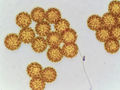
Under the microscope with a x40 objective, the tissues of the cup fungus Plicaria carbonaria collected near Pole Creek on North Wilderness Trail of Little Lake Creek Wilderness in Sam Houston National Forest north from Montgomery, Texas, reveal intricate details. The sample was collected on August 31, 2022. – generated by AI |
The image presents a microscopic view of tissues from the cup fungus Plicaria carbonaria, captured using an x40 objective lens. The image is dominated by the reddish-orange coloration of the fungal tissue, which is visible as irregularly shaped masses and cells. The tissue appears to be densely packed with fungal hyphae, which are thread-like structures that make up the body of the fungus. In addition to the reddish-orange coloration, there are also several smaller, darker-colored objects present in the image. These may represent spores or other reproductive structures of the fungus. The background of the image is a light brown color, which suggests that it is a microscope slide. The overall appearance of the image suggests that it was taken during a microscopic examination of a sample of Plicaria carbonaria tissue. Overall, the image provides a detailed view of the structure and organization of the fungal tissue, highlighting its unique characteristics and features. – AI vision |
Los tejidos del hongo en copa Plicaria carbonaria bajo un microscopio con objetivo x40, recolectados cerca de Pole Creek en el North Wilderness Trail de Little Lake Creek Wilderness en Sam Houston National Forest al norte de Montgomery. Texas, 31 de agosto de 2022. La imagen presenta una vista microscópica de los tejidos del hongo en copa Plicaria carbonaria, capturada usando un objetivo x40. La imagen está dominada por la coloración rojiza-naranja del tejido fúngico, visible como masas y células de forma irregular. El tejido parece estar densamente empaquetado con hifas fúngicas, que son estructuras filamentosas que forman el cuerpo del hongo. Además de la coloración rojiza-naranja, también hay varios objetos más oscuros presentes en la imagen. Estos pueden representar esporas u otras estructuras reproductivas del hongo. El fondo de la imagen tiene un color marrón claro, lo que sugiere que se trata de un portaobjetos de microscopio. La apariencia general de la imagen sugiere que fue tomada durante un examen microscópico de una muestra de tejido de Plicaria carbonaria. En general, la imagen proporciona una vista detallada de la estructura y organización del tejido fúngico, destacando sus características únicas y peculiaridades. – Spanish translation |
Keyword variants
microscopy
micrograph
Lake Creek Wilderness visual image
high resolution online digital collection
North Wilderness Trail pics
Houston National Forest image
Lone Star State
Little Lake photos
United States
picture gallery
photo album
documentary photographs
daily pics
TX
travelogue
snapshots
sightseeing
photowalks
photoblog
littel
graphic
fotografias
foto
America