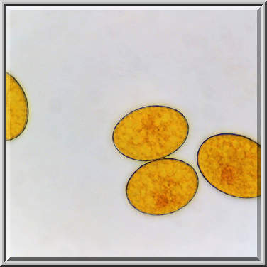
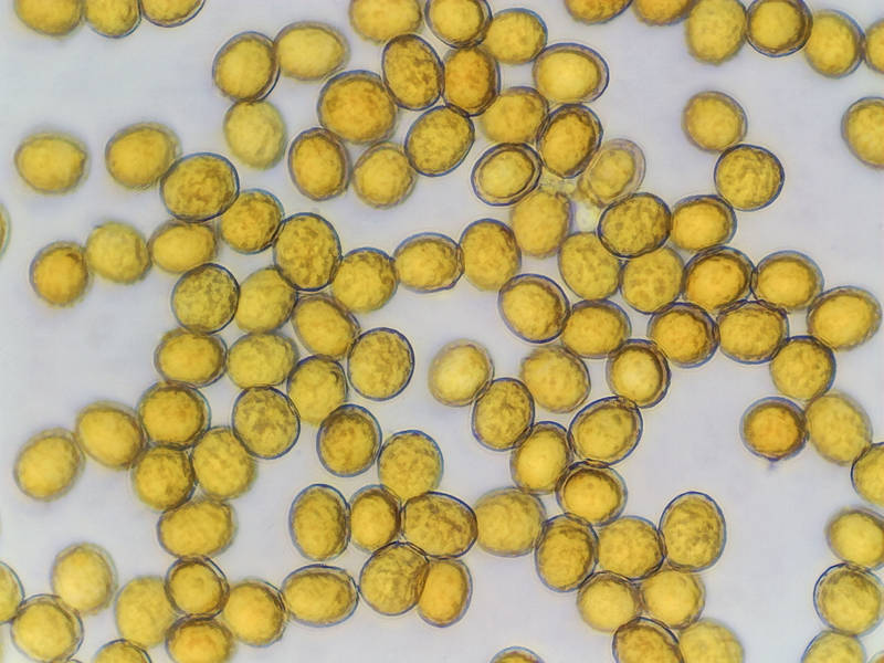
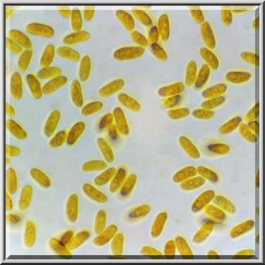
Spores of light brown corticioid (crust) fungus from an oak or pecan log collected in Lick Creek ParkLick Creek: Background Notes a day before. College Station, Texas


Album 3333: College Station, Texas, January 22 - 24, 2023
The image shows spores of a light-brown corticioid fungus from an oak or pecan log that was collected in Lick Creek Park on January 23rd, 2023. The spores are numerous and densely packed within the image frame.
Spores are typically microscopic and spherical in shape, often appearing as small, round dots under a microscope. They can range in size from a few micrometers to several hundred micrometers in diameter, depending on the species.
In this case, there is no visible detail of the spores' surface features, which could include texture, color patterns, or other characteristics that would aid in identifying the specific fungus species.
The background of the image appears to be a light-colored surface, possibly a microscope slide or petri dish. The spores are scattered across this surface, with some appearing to be overlapping or touching one another.
Overall, the image provides a general visual representation of the spores from the corticioid fungus without offering detailed information about their size, shape, color, or other characteristics that could help identify the specific species. – AI vision
🇪🇸 Descripción en español / Spanish Description
Las esporas de un hongo corticioso pardo claro (costra) recolectado de un tronco de roble o pacana el día anterior en Lick Creek Park, College Station, Texas, 24 de enero del 2023. La imagen muestra esporas de un hongo corticioso pardo claro de un tronco de roble o pacana recolectado en Lick Creek Park el 23 de enero del 2023. Las esporas son numerosas y densamente empaquetadas dentro del marco de la imagen. Las esporas suelen ser microscópicas y de forma esférica, a menudo aparecen como pequeños puntos redondos bajo un microscopio. Su tamaño puede variar desde unos pocos micrómetros hasta varios cientos de micrómetros de diámetro, dependiendo de la especie. En este caso, no se observan detalles visibles de las características superficiales de las esporas, que podrían incluir textura, patrones de color u otras características que ayudarían a identificar la especie de hongo específica. El fondo de la imagen parece ser una superficie de color claro, posiblemente un portaobjetos o placa Petri. Las esporas están dispersas en esta superficie, algunas de ellas solapadas o tocándose entre sí. En general, la imagen proporciona una representación visual general de las esporas del hongo corticioso sin ofrecer información detallada sobre su tamaño, forma, color u otras características que podrían ayudar a identificar la especie específica. – Spanish translation


