 |
| |||||||||||||||

| ||||||||||||||||


| Similar |   |
  |
  |
  |
  |
| |||||||||||||||||||||||||||
| |||||||||||||||||||||||||||
| 1996 1997 1998 1999 2000 2001 2002 2003 2004 2005 2006 2007 2008 2009 2010 2011 2012 2013 2014 2015 2016 2017 2018 2019 2020 2021 2022 2023 2024 2025 2026 | |||||||||||||||||||||||||||
| New version Slide Show Most Popular Full List Statistics Technical Details Home Page | |||||||||||||||||||||||||||
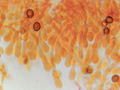
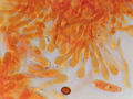
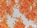
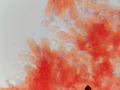
The orange oysterling mushrooms were observed to have basidia and spores on a vertically cut surface of an oak log in Sam Houston National Forest, located north of Montgomery, Texas. The observation was made on November 12, 2023. – generated by AI |
The image displays a microscopic view of the gills of an orange oysterling mushroom (Pleurotus levis), likely found on a vertically cut surface of an oak log in Sam Houston National Forest, Texas. The sample was collected by a mycologist and photographed through a microscope at 40x magnification. The image features elongated basidia, which are spore-bearing structures that resemble tiny stalks with bulbous bases. The spores are arranged along the length of each basidium, resembling small dots on a string. Two orange-colored spores appear to be located near the top left side of the image, while several more can be seen scattered throughout. The background is out of focus and appears white or slightly grayish in color. The image's resolution is sufficient for identifying the mushroom species but may not provide enough detail for further analysis without additional magnification or specialized equipment. Overall, the image effectively captures the microscopic details of a mushroom's gills, providing valuable information for mycological research and identification purposes. – AI vision |
Los basidios y esporas de los hongos oysterling naranjas en una superficie cortada verticalmente de un tronco de roble, recolectado en el Bosque Nacional Sam Houston al norte de Montgomery, Texas, el 12 de noviembre de 2023. La imagen muestra una vista microscópica de las láminas de un hongo oysterling naranja (Pleurotus levis), que probablemente se encontraba en una superficie cortada verticalmente de un tronco de roble en el Bosque Nacional Sam Houston, Texas. La muestra fue recolectada por un micólogo y fotografiada a través de un microscopio con un aumento de 40x. La imagen presenta basidios alargados, que son estructuras portadoras de esporas que se asemejan a pequeños tallos con bases bulbosas. Las esporas están dispuestas a lo largo del longitud del basidio, pareciendo pequeños puntos en una cadena. Dos esporas de color naranja aparecen ubicadas cerca de la esquina superior izquierda de la imagen, mientras que otras más se pueden ver dispersas por toda ella. El fondo está desenfocado y aparece de color blanco o ligeramente grisáceo. La resolución de la imagen es suficiente para identificar la especie de hongo pero puede no proporcionar detalles suficientes para un análisis adicional sin aumentos adicionales o equipamiento especializado. En general, la imagen captura efectivamente los detalles microscópicos de las láminas de un hongo, proporcionando información valiosa para fines de investigación y identificación micológica. – Spanish translation |
Keyword variants
microscopy
micrograph
high resolution online digital collection
Houston National Forest picture
Lone Star State
United States
picture gallery
photo album
documentary photographs
daily pics
TX
travelogue
snapshots
sightseeing
photowalks
photoblog
graphic
fungus
fungi
fotografias
foto
America