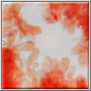
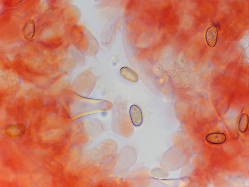
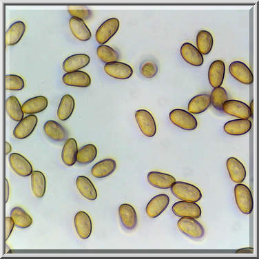
Tissues (in Congo Red) of chestnut brittlestem mushrooms Homophron spadiceum (Psathyrella spadicea) from the base of a hackberry tree, collected in Lick Creek ParkLick Creek: Background Notes. College Station, Texas


Album 3426: College Station, Texas, November 15 - 16, 2023
The image depicts a microscopic view of fungal tissue, likely obtained through microscopy or photomicrography, using Congo Red stain. The background appears to be orange due to the staining process; however, this coloration is not representative of the actual organism's natural colors.
Upon closer inspection, several elongated cells with dark blue rings surrounding them are visible. These cells may be hyphae, which are characteristic of fungi and other related organisms. They often appear as long, thin filaments under a microscope.
The image also features numerous smaller, irregularly shaped structures that could represent spores or conidia. Spores in fungal tissue can vary significantly in size and shape depending on the species; therefore, without more detailed information about the specific organism being examined, it is difficult to determine their exact nature.
Further details are required to accurately identify these microscopic features; however, based solely on this image, they appear to be components of a fungus's reproductive structures. – AI vision
🇪🇸 Descripción en español / Spanish Description
Tejidos (en rojo Congo) de hongos Homophron spadiceum (Psathyrella spadicea) de castaño delicuescente recogidos en el Parque Lick Creek. College Station, Texas, 15 de noviembre de 2023. La imagen muestra una vista microscópica del tejido fúngico, probablemente obtenida mediante microscopía o fotomicrografía, con tinción rojo Congo. El fondo aparece naranja debido al proceso de tinción; sin embargo, esta coloración no es representativa de los colores naturales reales del organismo. Tras un examen más detallado, son visibles varias células alargadas con anillos azules oscuros que las rodean. Estas células pueden ser hifas, que son características de hongos y otros organismos relacionados. Con frecuencia aparecen como filamentos largos y delgados bajo un microscopio. La imagen también presenta numerosas estructuras más pequeñas y con formas irregulares que podrían representar esporas o conidios. Las esporas en el tejido fúngico pueden variar significativamente de tamaño y forma dependiendo de la especie; por lo tanto, sin información más detallada sobre el organismo específico examinado, es difícil determinar su exacta naturaleza. Se requieren datos adicionales para identificar con precisión estas características microscópicas; sin embargo, basándonos únicamente en esta imagen, parecen ser componentes de las estructuras reproductivas de un hongo. – Spanish translation


