
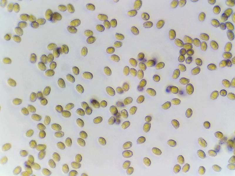

Spores of light brown corticioid tooth fungus on an oak(?) log, collected two days before in Hensel Park. College Station, Texas


Album 3428: Sam Houston Forest, Texas, November 18, 2023
The image presents a microscopic view of spores from the corticioid tooth fungus, which was previously observed on an oak log at Hensel Park in College Station, Texas. The spores are characterized by their light brown hue and oval shape. They exhibit varying sizes, with most being approximately 3 to 5 micrometers (μm) in length.
The spore coloration can be attributed to the presence of melanin or other pigments within the fungal cell wall. These pigments play a crucial role in protecting the fungus against environmental stressors and may also contribute to its ability to absorb nutrients from its surroundings.
In terms of their shape, the oval form is typical for corticioid fungi, which are known for their characteristic "tooth-like" or "bracket-like" fruiting bodies. This morphology allows them to efficiently capture and digest organic matter in their environment.
The size variation among the spores can be attributed to factors such as genetic diversity within the fungal population, environmental influences during sporulation, or differences in nutrient availability that impact growth rates. It is important to note that these variations do not necessarily reflect any inherent characteristics of the fungus itself but rather its adaptation to specific conditions at the time of sampling.
Overall, this image provides valuable insights into the microscopic features and potential adaptations of the corticioid tooth fungus, highlighting its unique combination of light brown coloration and oval-shaped spores. – AI vision
🇪🇸 Descripción en español / Spanish Description
Las esporas del hongo dentario corticial de marrón claro en un tronco de roble (?), recolectadas dos días antes en Hensel Park. College Station, Texas, 18 de noviembre de 2023. La imagen presenta una vista microscópica de las esporas del hongo dentario corticial, que se observó previamente en un tronco de roble en Hensel Park en College Station, Texas. Las esporas se caracterizan por su marrón claro y forma ovalada. Presentan tamaños variables, con la mayoría siendo de aproximadamente 3 a 5 micrómetros (μm) de longitud. La coloración de las esporas puede atribuirse a la presencia de melanina u otros pigmentos dentro de la pared celular del hongo. Estos pigmentos desempeñan un papel crucial en la protección del hongo contra los factores estresantes del medio ambiente y también pueden contribuir a su capacidad para absorber nutrientes de sus alrededores. En términos de su forma, la forma ovalada es típica para los hongos corticiales, que se conocen por sus característicos cuerpos fructíferos "dentados" o "de corchete". Esta morfología les permite capturar y digerir eficientemente la materia orgánica en su entorno. La variación de tamaño entre las esporas puede atribuirse a factores como la diversidad genética dentro de la población fúngica, las influencias ambientales durante la esporulación o las diferencias en la disponibilidad de nutrientes que afectan las tasas de crecimiento. Es importante destacar que estas variaciones no necesariamente reflejan ninguna característica inherente del hongo en sí, sino su adaptación a condiciones específicas en el memento del muestreo. En general, esta imagen proporciona información valiosa sobre las características microscópicas y potenciales adaptaciones del hongo dentario corticial, destacando su combinación única de coloración marrón claro y esporas de forma ovalada. – Spanish translation


