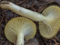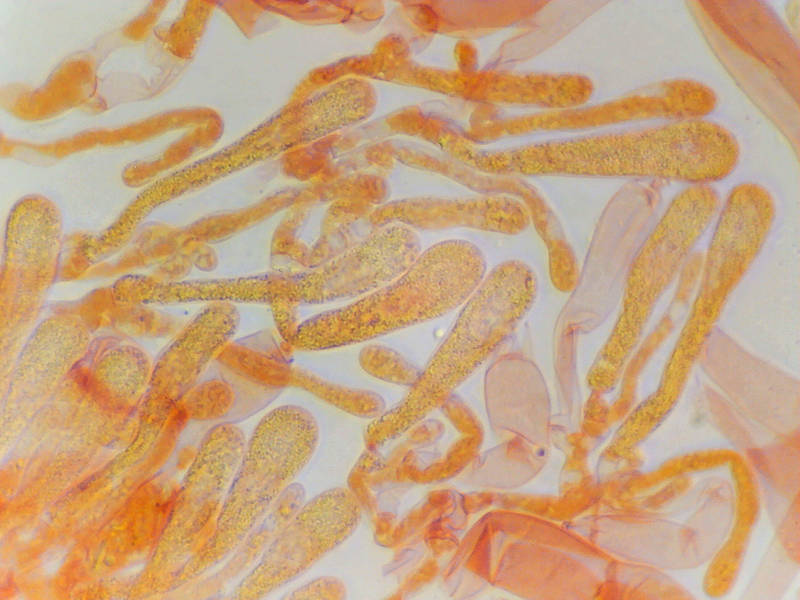

Cystidia and basidia of mushrooms Hygrophorus hypothejus(?) collected in Sam Houston National Forest near Montgomery. Texas


Album 3456: Sam Houston Forest, Texas, January 6, 2024
The image shows a microscopic view of the cystidia and basidia from a mushroom species identified as *Hygrophorus hypothejus*, although there is some ambiguity regarding the specific identification due to the lack of clear details in the caption.
**Observations:**
* The image displays a collection of elongated, rod-shaped structures with pointed ends.
* These structures are likely cystidia, which are sterile cells found on the surface of mushrooms and involved in various functions such as defense mechanisms or water absorption.
* In addition to the cystidia, there are also visible basidia present in the image. Basidia are specialized hyphal cells responsible for producing spores during the sexual reproduction cycle of fungi.
**Key Features:**
* **Cystidia:** The dominant structures in the image, these elongated cells have pointed ends and are scattered throughout the field.
* **Basidia:** These are also visible but less prominent than the cystidia. They appear as slightly larger, more rounded structures amidst the cystidia.
**Conclusion:**
Based on the information provided and the visual characteristics observed in the image, it is reasonable to conclude that the specimen depicted belongs to a species of mushroom within the genus *Hygrophorus*. The presence of both cystidia and basidia suggests that this fungus undergoes sexual reproduction, releasing spores from the basidia for dispersal.
**Further Identification:**
To confirm the exact species identification beyond *Hygrophorus hypothejus*, further microscopic examination or molecular analysis might be necessary. This could involve studying additional features such as the shape and size of the cystidia and basidia, the arrangement of these structures on the mushroom's surface, or conducting DNA sequencing to identify specific genetic markers unique to this species.
In summary, while the image provides valuable insights into the microscopic anatomy of a mushroom from the genus *Hygrophorus*, a more precise identification would require additional detailed examination and analysis. – AI vision
🇪🇸 Descripción en español / Spanish Description
Traduzca el siguiente texto del inglés al español:
Cystidia y basidios de hongos Hygrophorus hypothejus(?) recogidos en la Sam Houston National Forest cerca de Montgomery, Texas, el 6 de enero de 2024. La imagen muestra una vista microscópica de los cystidia y basidios de un hongo identificado como *Hygrophorus hypothejus*, aunque hay alguna ambigüedad en cuanto a la identificación específica debido a la falta de detalles claros en el pie de foto. **Observaciones:** * La imagen muestra una colección de estructuras alargadas, en forma de varilla con extremos puntiagudos. * Estas estructuras son probablemente cystidia, que son células estériles encontradas en la superficie de los hongos y participan en diversas funciones como mecanismos de defensa o absorción de agua. * Además de los cystidia, también hay basidios presentes en la imagen. Los basidios son células de hifas especializadas responsables de producir esporas durante el ciclo de reproducción sexual de los hongos. **Características clave:** * **Cystidia:** Las estructuras dominantes en la imagen, estas células alargadas tienen extremos puntiagudos y están dispersas por todo el campo. * **Basidia:** También son visibles pero menos prominentes que los cystidia. Aparecen como estructuras ligeramente más grandes y redondeadas entre las cystidia. **Conclusión:** Basándose en la información proporcionada y las características visuales observadas en la imagen, es razonable concluir que el espécimen representado pertenece a una especie de hongo dentro del género *Hygrophorus*. La presencia de cystidia y basidios sugiere que este hongo experimenta reproducción sexual, liberando esporas desde los basidios para su dispersión. **Identificación adicional:** Para confirmar la identificación específica más allá de *Hygrophorus hypothejus*, puede ser necesaria una examen microscópico adicional o un análisis molecular. Esto podría implicar estudiar características adicionales como la forma y el tamaño de los cystidia y basidios, la disposición de estas estructuras en la superficie del hongo o realizar una secuenciación de ADN para identificar marcadores genéticos únicos para esta especie. En resumen, aunque la imagen proporciona valiosas informaciones sobre la anatomía microscópica de un hongo del género *Hygrophorus*, una identificación más precisa requeriría un examen y análisis detallados adicionales. – Spanish translation