
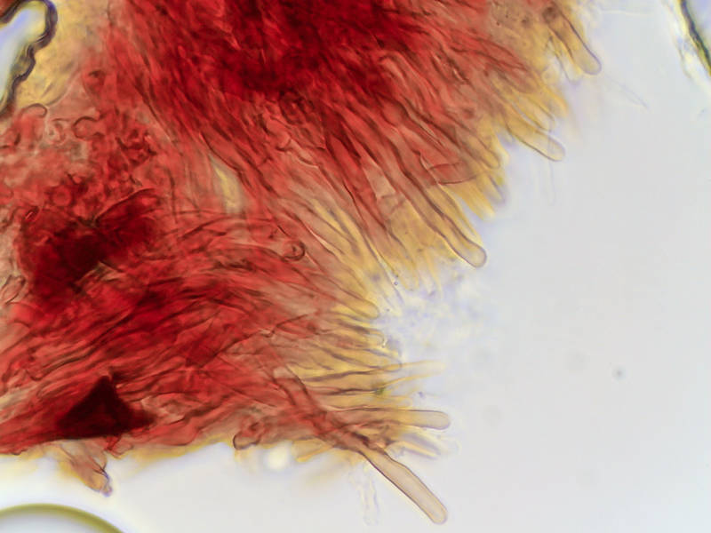

Hyphae (in KOH) of thin violet and brown corticioid fungus from a fallen honey locust, collected in Lick Creek ParkLick Creek: Background Notes. College Station, Texas


Album 3474: Sam Houston Forest - College Station, Texas, February 18 - 20, 2024
The image shows hyphae of a thin violet and brown corticioid fungus from a fallen honey locust tree, collected in Lick Creek Park, College Station, Texas on February 20, 2024.
Hyphae are the branching filaments that make up the mycelium (vegetative part) of fungi. The hyphae in this image appear as thin, elongated structures with a smooth surface. They are colored violet and brown and display varying thicknesses. Some hyphae appear thin and fragile while others appear thicker and more robust.
The hyphae are arranged in an irregular pattern, with some growing straight out from the center of the image and others branching off at an angle. The background of the image is white, providing a clean contrast to the colorful hyphae.
Overall, the image provides a detailed view of the structure and morphology of the fungus's mycelium, highlighting its unique characteristics and growth patterns. – AI vision
🇪🇸 Descripción en español / Spanish Description
Las hifas (en KOH) de un hongo corticioso fino morado y marrón recogido de una gomera caída, recolectadas en el Lick Creek Park. College Station, Texas, el 20 de febrero del 2024. La imagen muestra las hifas de un fino hongo corticioso morado y marrón de una gomera caída, recogido en el Lick Creek Park, College Station, Texas el 20 de febrero del 2024. Las hifas son los filamentos ramificados que forman el micelio (parte vegetativa) de los hongos. Las hifas en esta imagen aparecen como estructuras alargadas y delgadas con una superficie lisa. Están coloreadas de morado y marrón y presentan grosores variables. Algunas hifas parecen delgadas y frágiles, mientras que otras son más gruesas y robustas. Las hifas están dispuestas en un patrón irregular, con algunas creciendo directamente desde el centro de la imagen y otras ramificándose formando ángulos. El fondo de la imagen es blanco, proporcionando un contraste limpio a las hifas coloridas. En general, la imagen proporciona una vista detallada de la estructura y morfología del micelio del hongo, destacando sus características únicas y patrones de crecimiento. – Spanish translation


