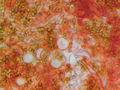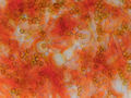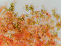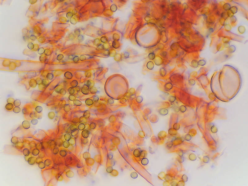

Tissues of light violet fluffy corticioid fungus from on old polypore, collected in Lick Creek ParkLick Creek: Background Notes. College Station, Texas


Album 3476: College Station, Texas, February 23, 2024
The image presents a microscopic view of fungal hyphae and spores, which are characteristic features of fungi. The overall appearance suggests that the fungus is likely a type of Ascomycota or Basidiomycota, given its fluffy corticioid growth habit.
**Key Features:**
* **Hyphae:** The image shows numerous hyphae with varying diameters and shapes, indicating different stages of development.
* Hyphae are branching filaments that make up the mycelium of fungi.
* They play a crucial role in nutrient uptake and transportation within the fungal body.
* **Spores:** Various spore types are visible throughout the image, including spherical, oval, and elongated shapes.
* Spores are reproductive structures responsible for dispersing fungi to new environments.
* Their diversity in shape and size may indicate different functions or adaptations for survival.
* **Coloration:** The hyphae exhibit a light violet hue, while the spores appear in shades of brown, orange, yellow, green, and blue.
* Coloration can provide clues about the fungus's pigmentation patterns and possibly its ecological niche.
**Conclusion:**
Based on the microscopic characteristics observed, it is reasonable to infer that the fungus belongs to a species within the Ascomycota or Basidiomycota phyla. Further analysis of the spore morphology, size distribution, and any distinctive structures such as perithecia (fruiting bodies) would be necessary for a definitive identification at the species level.
**Recommendation:**
For more precise classification, it is recommended to examine additional microscopic features, such as:
* **Spore wall structure:** The presence of ornamentations or appendages could help narrow down the taxonomy.
* **Fruit body morphology:** If present, examining the shape, size, and arrangement of fruit bodies (e.g., perithecia) would aid in identification.
* **Cultural characteristics:** Growing the fungus in a controlled environment may reveal additional features like mycelial growth patterns or pigment production.
By considering these factors, researchers can better understand the biology and ecology of this particular fungus species. – AI vision
🇪🇸 Descripción en español / Spanish Description
Los tejidos de un hongo corticioide esponjoso de color violeta claro recolectado en un poliporo viejo en Lick Creek Park, College Station, Texas, el 23 de febrero del 2024. La imagen presenta una vista microscópica de las hifas y esporas fúngicas, que son características comunes en los hongos. El aspecto general sugiere que el hongo es probablemente un tipo de Ascomicetes o Basidiomicetes, dado su hábitat corticioide esponjoso.
**Características clave:**
* **Hifas:** La imagen muestra numerosas hifas con diámetros y formas variables, lo que indica diferentes etapas de desarrollo.
* Las hifas son filamentos ramificados que componen el micelio de los hongos.
* Desempeñan un papel fundamental en la absorción de nutrientes y su transporte dentro del cuerpo fúngico.
* **Es poros:** Son visibles diversos tipos de esporas en toda la imagen, incluyendo formas esféricas, ovaladas y alargadas.
* Las esporas son estructuras reproductivas responsables de dispersar los hongos a nuevos entornos.
* Su diversidad en forma y tamaño puede indicar diferentes funciones o adaptaciones para la supervivencia.
* **Coloración:** Las hifas presentan un matiz violeta claro, mientras que las esporas aparecen en tonalidades marrones, naranjas, amarillas, verdes y azules.
* La coloración puede proporcionar pistas sobre los patrones de pigmentación del hongo y posiblemente su nicho ecológico.
**Conclusión:** En base a las características microscópicas observadas, es razonable inferir que el hongo pertenece a una especie dentro de los filos Ascomicetes o Basidiomicetes. Sería necesario un análisis adicional de la morfología de las esporas, la distribución del tamaño y cualquier estructura distintiva tal como peritecios (cuerpos fructíferos) para una identificación definitiva a nivel de especie.
**Recomendación:** Para una clasificación más precisa, se recomienda examinar características microscópicas adicionales, tales como:
* **Estructura de la pared de las esporas:** La presencia de ornamentaciones o apéndices podría ayudar a reducir la taxonomía.
* **Morfología del cuerpo fructífero:** Si están presentes, el examen de la forma, tamaño y disposición de los cuerpos fructíferos (por ejemplo, peritecios) ayudaría a la identificación.
* **Características culturales:** El cultivo del hongo en un entorno controlado puede revelar características adicionales como patrones de crecimiento micelial o producción de pigmentos.
Al considerar estos factores, los investigadores pueden entender mejor la biología y ecología de esta especie particular de hongos. – Spanish translation