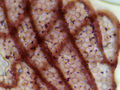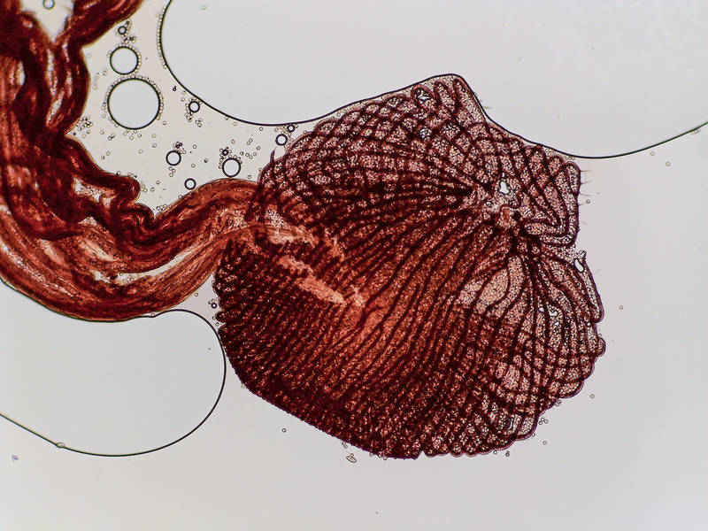
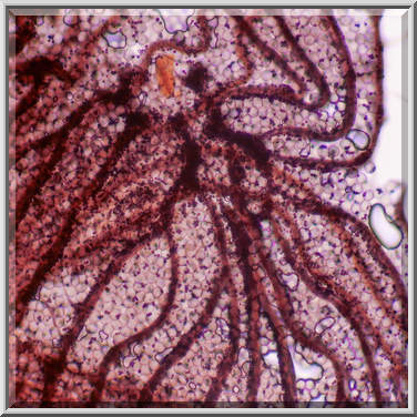
Stalked sporangia of slime mold Physarum viride under a microscope at lowest magnification, collected from brown rotten bark on oak log on Caney Creek Trail (Little Lake Creek Loop Trail) in Sam Houston National Forest north from Montgomery. Texas


Album 3150: College Station - Sam Houston Forest, Texas, May 9 - 12, 2022
The image is a photograph of a microscope slide containing an organism called Physarum viride, also known as yellow mycetozoa or common plasmodial slime mold.
Physarum viride is characterized by its bright yellow color and the presence of stalked sporangia.
The sporangium is attached to the substrate through a long stalk that provides support and protection for the spores inside. The sporangium contains numerous small, round, brownish-black spores that are released into the air when conditions become favorable for germination.
When viewed under a microscope at low magnification (about 40x), the sporangia appear as large, spherical structures with distinct cell walls and membranes. They have a smooth surface and lack any visible appendages or features.
The stalk supporting each sporangium is long and slender, often exceeding several millimeters in length. It tapers to a thin tip at the base of the sporangium, where it attaches firmly to the substrate.
Physarum viride typically grows on decaying plant material such as logs or tree bark. Its bright yellow coloration makes it easily recognizable among other organisms found in these environments.
Overall, the image provides an excellent visual representation of Physarum viride's unique morphology and helps identify this fascinating species within its natural habitat. – AI vision
🇪🇸 Descripción en español / Spanish Description
Stalked esporangios del hongo físico Physarum viride bajo un microscopio con el menor aumento posible, recogidos de la corteza podrida marronosa en un tronco de roble en el Caney Creek Trail (Little Lake Creek Loop Trail) en el Bosque Nacional Sam Houston al norte de Montgomery. Texas, 12 de mayo de 2022. La imagen es una fotografía de un portaobjetos de microscopio que contiene un organismo llamado Physarum viride, también conocido como micetozoos amarillos o moho común de esporas plasmodiales. Physarum viride se caracteriza por su color amarillo brillante y la presencia de esporangios con pedúnculo. El esporango está unido al sustrato a través de un largo tallo que proporciona soporte y protección para las esporas dentro. El esporango contiene numerosas esporas pequeñas, redondas y parduzcas que se liberan al aire cuando las condiciones son favorables para la germinación. Cuando se ven bajo un microscopio con poco aumento (aproximadamente 40x), los esporangios parecen ser estructuras esféricas grandes con distintas paredes y membranas celulares. Tienen una superficie lisa y carecen de apéndices o características visibles. El tallo que soporta cada esporango es largo y delgado, a menudo superando los varios milímetros de longitud. Se estrecha hacia una punta delgada en la base del esporango, donde se une firmemente al sustrato. Physarum viride normalmente crece en material vegetal en descomposición, como troncos o corteza de árboles. Su coloración amarilla brillante lo hace fácilmente reconocible entre otros organismos encontrados en estos entornos. En general, la imagen proporciona una excelente representación visual de la morfología única de Physarum viride y ayuda a identificar esta fascinante especie dentro de su hábitat natural. – Spanish translation