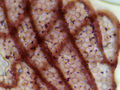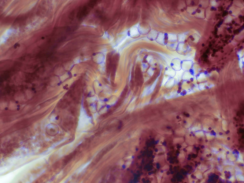

Tissues and spores of slime mold Physarum viride under a microscope, collected from an oak log on Caney Creek Trail (Little Lake Creek Loop Trail) in Sam Houston National Forest north from Montgomery. Texas


Album 3150: College Station - Sam Houston Forest, Texas, May 9 - 12, 2022
The image shows a microscopic view of tissues and spores of slime mold Physarum viride collected from an oak log on Caney Creek Trail (Little Lake Creek Loop Trail) in Sam Houston National Forest north from Montgomery, Texas on May 12, 2022.
The image is dominated by the pinkish-red color of the fungal tissue, which appears to be irregularly shaped and has a somewhat amorphous appearance. The spores are small and dark purple in color, scattered throughout the tissue. They appear to be spherical or oval-shaped and have a smooth surface.
In the background, there are some faint blue lines visible, which may represent the cell walls of the fungal cells. There is also a bright white area at the top left of the image, which could be due to lighting effects during microscopy.
Overall, the image provides a detailed view of the microscopic structure of Physarum viride, showcasing its unique features and characteristics. The presence of spores suggests that the fungus is in a reproductive stage, while the irregularly shaped tissue may indicate growth or development processes. – AI vision
🇪🇸 Descripción en español / Spanish Description
Los tejidos y esporas del liquenífago Physarum viride bajo un microscopio, recolectados de un tronco de roble en el Caney Creek Trail (Little Lake Creek Loop Trail) en el Bosque Nacional Sam Houston al norte de Montgomery. Texas, 12 de mayo de 2022. La imagen muestra una vista microscópica de tejidos y esporas del liquenífago Physarum viride recolectados de un tronco de roble en el Caney Creek Trail (Little Lake Creek Loop Trail) en el Bosque Nacional Sam Houston al norte de Montgomery, Texas el 12 de mayo de 2022. La imagen está dominada por el color rosa rojizo del tejido fungoso, que parece tener una forma irregular y un aspecto algo amorfo. Las esporas son pequeñas y de color púrpura oscuro, dispersadas en todo el tejido. Parecen serféricos u ovalados y tienen una superficie lisa. En segundo plano, se pueden ver líneas azules débiles, que podrían representar las paredes celulares de las células fúngicas. También hay una área blanca brillante en la esquina superior izquierda de la imagen, que podría deberse a efectos de iluminación durante la microscopía. En general, la imagen proporciona una vista detallada de la estructura microscópica de Physarum viride, mostrando sus características y rasgos únicos. La presencia de esporas sugiere que el hongo está en un estadio reproductivo, mientras que el tejido irregularmente conformado puede indicar procesos de crecimiento o desarrollo. – Spanish translation