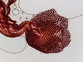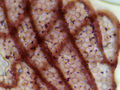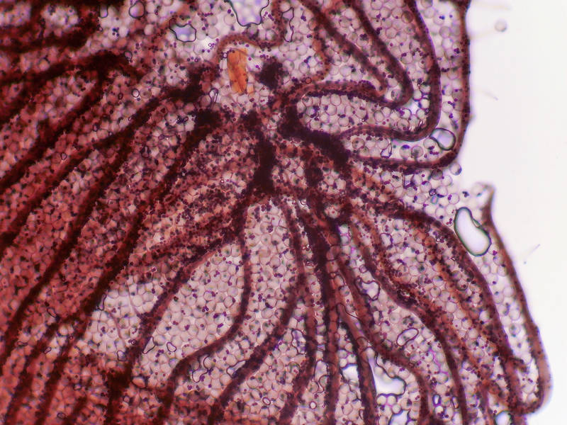

Slime mold Physarum viride under a microscope at low magnification, collected from an oak log on Caney Creek Trail (Little Lake Creek Loop Trail) in Sam Houston National Forest north from Montgomery. Texas


Album 3150: College Station - Sam Houston Forest, Texas, May 9 - 12, 2022
The image shows a microscopic view of slime mold Physarum viride at low magnification, captured on May 12, 2022, from an oak log on Caney Creek Trail (Little Lake Creek Loop Trail) in Sam Houston National Forest north from Montgomery, Texas.
**Physical Characteristics**
* The specimen is large and irregularly shaped, with a network of veins or tubes visible throughout.
* It has a translucent appearance, with a pinkish-red hue that may be due to the presence of pigments or other substances within the cells.
* The surface of the specimen appears rough and uneven, with small bumps or protrusions scattered across its surface.
**Cellular Structure**
* Upon closer inspection, it becomes clear that the specimen is composed of individual cells.
* These cells are connected by thin filaments or fibers, forming a network that gives the specimen its characteristic shape and structure.
* The cells themselves appear to be relatively large compared to other microscopic organisms, with some measuring up to several hundred micrometers in diameter.
**Overall Impression**
The image provides a fascinating glimpse into the world of slime molds, highlighting their unique cellular structure and ability to form complex networks. – AI vision
🇪🇸 Descripción en español / Spanish Description
El hongo slimy Physarum viride bajo un microscopio con baja ampliación, recolectado de una tronca de roble en el Caney Creek Trail (Little Lake Creek Loop Trail) en el Bosque Nacional Sam Houston al norte de Montgomery. Texas, 12 de mayo de 2022. La imagen muestra una vista microscópica del moho slimy Physarum viride con baja ampliación, capturada el 12 de mayo de 2022, de una tronca de roble en Caney Creek Trail (Little Lake Creek Loop Trail) en el Bosque Nacional Sam Houston al norte de Montgomery, Texas. **Características Físicas** * El espécimen es grande y de forma irregular, con una red de venas o tubos visibles a lo largo. * Tiene un aspecto traslúcido, con un tono rosado-rojizo que puede deberse a la presencia de pigmentos u otras sustancias dentro de las células. * La superficie del espécimen parece rugosa e irregular, con pequeños bultos o protuberancias dispersados en su superficie. **Estructura Celular** * Tras un examen más cercano, queda claro que el espécimen está compuesto por células individuales. * Estas células están conectadas por filamentos finos o fibras, formando una red que le da al espécimen su característica forma y estructura. * Las propias células parecen ser relativamente grandes en comparación con otros microorganismos, algunas miden hasta varios cientos de micrómetros de diámetro. **Impresión General** La imagen proporciona una interesante visión del mundo de los mohos slimy, resaltando su única estructura celular y capacidad para formar redes complejas. – Spanish translation