 |
| |||||||||||||||

| ||||||||||||||||


| Similar |   |
  |
  |
  |
  |
| |||||||||||||||||||||||||||
| |||||||||||||||||||||||||||
| 1996 1997 1998 1999 2000 2001 2002 2003 2004 2005 2006 2007 2008 2009 2010 2011 2012 2013 2014 2015 2016 2017 2018 2019 2020 2021 2022 2023 2024 2025 2026 | |||||||||||||||||||||||||||
| New version Slide Show Most Popular Full List Statistics Technical Details Home Page | |||||||||||||||||||||||||||
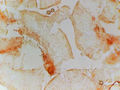
At a low magnification of x40, the tissues of the Ascomycete fungus Hyalorbilia inflatula appear as a mass of thin, branching filaments that are irregularly shaped and vary in size. The overall texture is rough and bumpy, with small protrusions and depressions covering the surface. The colors range from pale yellow to cream, with some areas exhibiting a faint pinkish hue. These tissues were collected on November 15, 2023, in Lick Creek Park, College Station, Texas. – generated by AI |
The image presents a microscopic view of fungal tissues, specifically those from the Ascomycete fungus Hyalorbilia inflatula. The specimen was collected on November 15th, 2023, at Lick Creek Park in College Station, Texas. **Image Characteristics:** * **Magnification:** The image is captured using a x40 objective lens. **Key Features:** * **Fungal Tissues:** The central focus of the image is the intricate structure of the fungal tissues. These appear as thin, branching filaments that are densely packed together. **Conclusion:** The provided image offers a detailed view of the microscopic structure of Hyalorbilia inflatula fungal tissues. This allows for observation of their intricate morphology and potential identification based on specific characteristics. – AI vision |
Los tejidos del hongo ascomiceto Hyalorbilia inflatula con bajo aumento (objetivo x40) recogido en Lick Creek Park. College Station, Texas, Noviembre 15, 2023. La imagen presenta una vista microscópica de los tejidos fúngicos, específicamente aquellos del hongo ascomiceto Hyalorbilia inflatula. El espécimen fue recolectado el 15 de noviembre de 2023 en Lick Creek Park en College Station, Texas. **Características de la imagen:** * **Aumento:** La imagen se captura utilizando un objetivo x40. * **Esquema de colores:** El fondo de la imagen presenta diferentes tonalidades de púrpura y gris, mientras que los tejidos fúngicos son principalmente blancos u off-white con algunos matices de marrón claro y rojo. * **Textura:** Los tejidos fúngicos muestran una delicada cualidad casi translúcida, con aristas y surcos visibles en su superficie. **Principales características:** * **Tejidos fúngicos:** El centro de la imagen es la estructura compleja de los tejidos fúngicos. Éstos aparecen como finos filamentos ramificados, densamente empaquetados juntos. * **Fondo:** El fondo de la imagen es relativamente sencillo, lo que sugiere que representa un medio transparente o una solución de montaje utilizada para preservar y visualizar la muestra. **Conclusión:** La imagen proporcionada ofrece una vista detallada de la estructura microscópica de los tejidos fúngicos Hyalorbilia inflatula. Esto permite la observación de su compleja morfología y la identificación potencial basada en características específicas. – Spanish translation |
Keyword variants
microscopy
micrograph
high resolution online digital collection
Lick Creek Park photos
Berlin on the Brazos
Rock Prairie Rd
Lone Star State
Greens Prairie Road
United States
picture gallery
photo album
nature trails
Nature Park
documentary photographs
daily pics
conservative town
Brazos County
wildlife
TX
travelogue
snapshots
sightseeing
photowalks
photoblog
graphic
fotografias
foto
Bryan
America
Aggieland