 |
| |||||||||||||||

| ||||||||||||||||


| Similar |   |
  |
  |
  |
  |
| |||||||||||||||||||||||||||
| |||||||||||||||||||||||||||
| 1996 1997 1998 1999 2000 2001 2002 2003 2004 2005 2006 2007 2008 2009 2010 2011 2012 2013 2014 2015 2016 2017 2018 2019 2020 2021 2022 2023 2024 2025 2026 | |||||||||||||||||||||||||||
| New version Slide Show Most Popular Full List Statistics Technical Details Home Page | |||||||||||||||||||||||||||
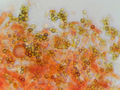
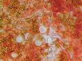
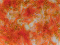
The image shows a microscopic view of fungal hyphae and spores, which are characteristic features of fungi. The presence of these structures suggests that the sample is likely from a fungus, specifically a corticioid species based on the description in the caption. **Key Features:** * **Hyphae:** The image reveals numerous thin, branching filaments that are typical of fungal hyphae. These filaments appear to be densely packed and intertwine with one another. **Coloration:** * The background of the image is predominantly white or light-colored, which could indicate the presence of a medium used in microscopy to enhance contrast and visibility. **Conclusion:** The provided image is likely a microscopic view of a fungal specimen, specifically a corticioid species identified based on the description in the caption. The presence of hyphae and spores supports this conclusion, as these structures are fundamental components of fungi. – AI vision |
Las hifas y las esporas de un hongo corticoides violeta claro peludo en un poliporo antiguo, recogido en Lick Creek Park. College Station, Texas, 23 de febrero del 2024. La imagen muestra una vista microscópica de las hifas y esporas fúngicas, que son características de los hongos. La presencia de estas estructuras sugiere que la muestra proviene probablemente de un hongo, específicamente de una especie corticoides basándose en la descripción en el subtítulo. **Características principales:** * **Hifas:** La imagen revela numerosos filamentos delgados y ramificados que son típicos de las hifas fúngicas. Estos filamentos parecen estar densamente empaquetados e interconectados entre sí. * **Especias:** Escatimadas a través de la imagen se encuentran pequeñas estructuras esféricas que se asemejan a esporas. Las esporas son unidades reproductivas de hongos responsables de la dispersión. **Coloración:** * El fondo de la imagen es predominantemente blanco o de color claro, lo que podría indicar la presencia de un medio usado en microscopía para mejorar el contraste y visibilidad. * Las hifas y las esporas presentan una gama de colores, incluyendo tonos de marrón, naranja y amarillo. Este matiz puede ser resultado de técnicas de tinción empleadas durante la preparación de la muestra. **Conclusión:** La imagen proporcionada es probablemente una vista microscópica de una muestra fúngica, específicamente una especie corticoides identificada en función de la descripción en el subtítulo. La presencia de hifas y esporas respalda esta conclusión, ya que estas estructuras son componentes fundamentales de los hongos. – Spanish translation |
Keyword variants
microscopy
micrograph
Lick Creek Park web graphics
high resolution online digital collection
Berlin on the Brazos
Rock Prairie Rd
Lone Star State
Greens Prairie Road
United States
picture gallery
photo album
nature trails
Nature Park
documentary photographs
daily pics
conservative town
Brazos County
wildlife
TX
travelogue
snapshots
sightseeing
photowalks
photoblog
graphic
fotografias
foto
Bryan
America
Aggieland